
Ribosomes, Golgi & Endoplasmic Reticulum
Compartmentalized Cell Structure
- Eukaryotic cells have a more complex ultrastructure than prokaryotic cells
- The cytoplasm of eukaryotic cells is divided up into membrane-bound compartments called organelles
- See notes on Topic 2.10 Cell Compartmentalization
Eukaryotic Animal Cell Structure Diagram

The ultrastructure of an animal cell shows a densely packed cell of compartmentalized organelles
Eukaryotic Plant Cell Structure Diagram
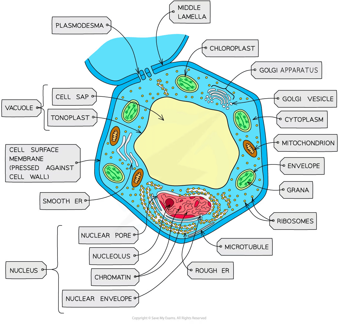
Plant cells have a larger, more regular structure in comparison to animal cells which also contains compartmentalized organelles
Ribosomes
- Ribosomes are found in all cells
- This fact supports the view that all life forms share a common ancestry
- Either freely in the cytoplasm (of all cells)...
- Or bound to the endoplasmic reticulum (ER) to form rough ER (only in eukaryotic cells)
- Ribosomes are the site of protein synthesis
- They consist of a large and a small subunit composed of protein and ribosomal RNA (rRNA)
- Protein provides structure to the ribosome
- rRNA facilitates the binding of mRNA and tRNA and catalyzes the formation of peptide bonds between amino acids
- Ribosomes have three tRNA binding sites and one mRNA binding site
- mRNA sits in a groove between the two subunits and the ribosome moves along, forming a polypeptide as it travels
The Structure of a Ribosome Diagram

A diagram of a ribosome, showing the small and large subunits and the strand of mRNA being translated
Free Ribosomes
- In eukaryotic cells, protein synthesis commonly occurs at free ribosomes in the cytoplasm
- Free ribosomes can move within the cytoplasm and synthesize proteins for use primarily within the cell
- As opposed to proteins destined to be secreted extracellularly
- Proteins synthesized on free ribosomes are destined for use within the cytosol (the fluid part of the cytoplasm)
- And within large organelles such as mitochondria and chloroplasts
Membrane-Bound Ribosomes
- When free ribosomes make proteins destined for lysosomes, or secretion from the cell, the ribosome becomes bound to the endoplasmic reticulum (ER)
- The synthesized protein can be carried via a vesicle to the Golgi apparatus before being secreted out of the cell
Endoplasmic reticulum
- Endoplasmic reticulum (ER) is a series of membrane-bound channels in the cytoplasm of eukaryotic cells
The Types of Endoplasmic Reticulum Diagram

The RER and ER are visible under the electron microscope
The presence or absence of ribosomes helps to tell them apart
Rough Endoplasmic Reticulum (RER)
- Its surface is studded with ribosomes
- Formed from continuous folds of membrane continuous with the nuclear envelope
- Its role is to process proteins made by the ribosomes
Smooth Endoplasmic Reticulum (ER)
- Does not have ribosomes on the surface; its function is distinct to the RER
- ER is involved in the production, processing and storage of lipids, carbohydrates and steroids
Golgi Apparatus

The structure of the Golgi apparatus
- Flattened sacs of membrane (called cisternae) are similar to the smooth endoplasmic reticulum
- The interior part of each cisterna, the lumen, holds the necessary enzymes for the Golgi to function
- The Golgi modifies proteins and lipids before packaging them into Golgi vesicles
- The vesicles then transport the proteins and lipids to their required destinations
- Proteins that go through the Golgi apparatus are usually
- Exported (eg, hormones such as insulin)
- Put into lysosomes (such as hydrolytic enzymes)
- Delivered to membrane-bound organelles
- These processes are commonly referred to as protein trafficking
Exam Tip
You will not need to learn the specific functions of smooth ER in certain specialized cells
Similarly, knowledge of the role of the Golgi in the synthesis of specific phospholipids, the packaging of specific enzymes for lysosomes, peroxisomes, and secretory vesicles is not required in the AP course description.
